




Introduction
More patients are undergoing placement of intracranial neurologic stimulators such as deep brain stimulators (DBS). The placement of a DBS is a neurosurgical procedure in which electrodes are placed in precise areas of the brain such as the subthalamic nucleus and the globus pallidus to treat conditions such as Parkinson’s, essential tremor, and obsessive-compulsive disorder. It is estimated that the number of new patients receiving a DBS is increasing by more than 12,000 per year worldwide.1 DBS procedures account for over 5500 admissions annually in the United States.1
Intraoperative neuromonitoring (IONM) is used to detect injury to the nervous system during surgery and has become common during higher risk procedures including spine surgery.2 Two types of evoked potentials that are often employed during spine surgery are motor evoked potentials (MEPs) and somatosensory evoked potentials (SSEPs). Motor pathway monitoring is performed using transcranial stimulation of up to several hundred volts, with detection of evoked signals from electrodes in peripheral muscles. Sensory pathway monitoring is conducted in reverse, with stimulation at the peripheral nerves and detection of evoked signals via cranial electrodes. SSEPs require significantly less stimulation (typically 20-50 mA) compared to MEPs.3
We report a case where SSEP monitoring was successfully and safely used in a patient with bilateral DBS undergoing a cervical spine fusion.
Case Description
An 80-year-old female with a past medical history of Parkinson’s disease on carbidopa-levodopa with bilateral DBS presented for an anterior discectomy and fusion (ACDF) for cervical stenosis under general anesthesia with utilization of IONM. Her additional past medical history included obstructive sleep apnea, breast cancer in remission, and lumbar spondylolisthesis.
The DBS were placed ten years ago as her disease had progressed beyond the control of medical management. The patient had undergone battery replacement two years prior, and her devices were functioning properly as was documented by her neurologist with a recent interrogation of the device.
The neurosurgeon who originally placed and managed the DBS was unaware she was scheduled for spine surgery. The spine surgeon had not considered possible interactions between the DBS and the planned IONM including transcranial MEPs (tcMEPs). On the day of surgery, the anesthesiologist identified a potential concern regarding the risks of the prescribed surgical plan in the presence of a DBS system in this patient.
A multidisciplinary discussion occurred prior to the procedure with the neurosurgeon who placed the DBS, the orthopedic surgeon performing the ACDF, the neurologist interpreting the IONM, and the neuroanesthesiologist. Regarding the IONM, it was decided to proceed with the use of SSEPs, but to avoid using MEPs. The neurosurgeon who placed the DBS felt the risk of damage or reprogramming was too high, given the surgical location, to utilize MEPs. The spine surgeon decided that the risk of employing MEPs in this patient outweighed the benefits given the recommendations from the neurosurgeon. Prior to leaving the pre-operative area, the patient’s DBS was turned off (Figure 1A) with the use of her remote.
The case proceeded uneventfully with steady SSEP signals throughout. The patient’s DBS was turned back on prior to emergence and extubation. (Figure 1B) The DBS generator site was examined in the recovery unit and there were no signs of a thermal burn or damage to the generator. Her DBS appeared to be properly functioning given the absence of Parkinsonian symptoms. Her post operative period was uneventful and on follow up with her neurologist, her post operative interrogation revealed no change in battery life or lead impedance.
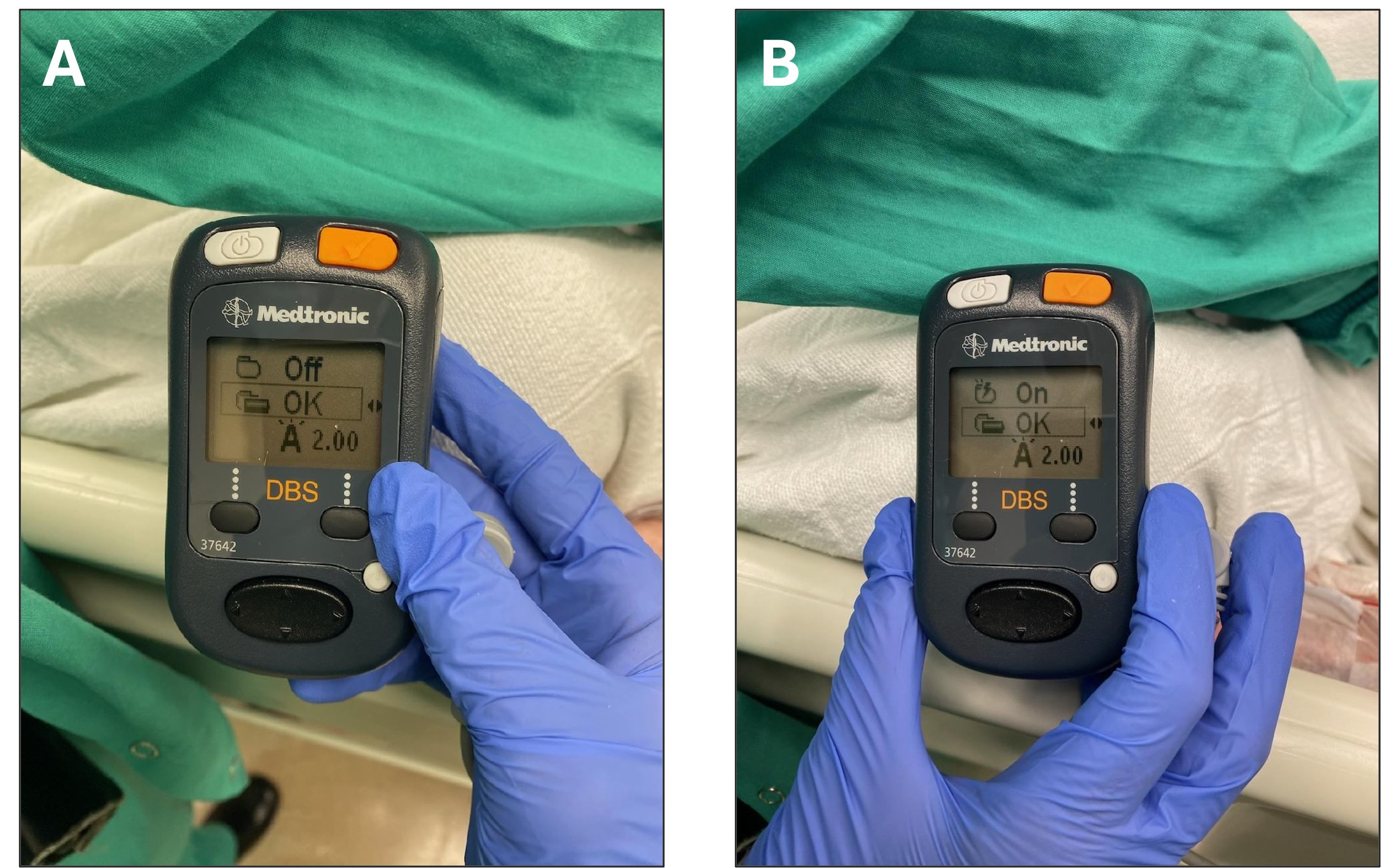
Figure 1. Image A shows the patient’s DBS stimulator in “off” mode, prior to induction. Image B shows the DBS in “on” mode prior to emergence and extubation.
Discussion
Patients with Parkinson’s Disease are now living longer and having more treatment options thanks to both advancements in medications and the use of intracranial neurologic stimulators. Surgical correction of the kyphoscoliosis and degenerative spondylosis that can accompany Parkinson’s Disease is crucial to improving quality of life for these patients. It is well documented that performing degenerative spine correction surgeries in this population is challenging; as many as one in three patients will need revision surgery.4 These surgeries are complicated in large part due to the muscular dysfunction and poor bone quality that patients with Parkinson’s Disease classically have,5 as well as the potential for interactions between their intracranial devices and standard IONM.
There is currently very little data to guide use of IONM in patients with intracranial implanted devices such as DBS. There are known risks to the device hardware and function, as well as to the patient’s tissues along the length of the device, when neuromonitoring is performed; these risks are currently not quantified. Given that no modality of neuromonitoring is completely free of risk, the IONM plan for patients with these devices should be individually tailored. It is imperative that these complex cases are preceded by a multidisciplinary discussion including the surgeon, anesthesiologist, neurologist, and neuromonitoring technician to ensure that all of the patient’s comorbidities have been considered, the surgical risks have been identified, and the surgical, anesthetic, and neuromonitoring plans are compatible with any implanted devices the patient may have. The patient should be included in the multidisciplinary discussion of the perioperative care plan. They should be made aware of the potentially increased risk associated with use of IONM in the setting of an implanted device, as well as the risk of omitting this monitoring. Additionally, should the decision be made to proceed with IONM, patients should be scheduled for both preoperative and postoperative visits with their neurologist in order to have their device interrogated, ensure proper functioning, and confirm appropriate lead impedance.
This case report aims to provide some evidence supporting the utilization of somatosensory neuromonitoring in patients with DBS. SSEPs represent an important modality for monitoring the integrity of the dorsal and lateral spinal cord intraoperatively. These signals are initiated in the periphery using electrodes placed along nerves in the patient’s extremities, travel along the dorsal column medial lemniscus pathway, and are ultimately recorded in the somatosensory cortex. The current required for these signals is very low, around 20-50mA, suggesting that the risk of damage to implanted devices should be relatively low. Additionally, avoiding their use for fear of interaction with implanted electrical devices exposes the patient to the potential risk of unrecognized intraoperative damage to the nervous system, which is not a negligible risk. The risk of neurologic damage has been reported at 0.5%-1.6% for scoliosis repairs6 and 0.4% for ACDF.7 Higher risk surgeries are associated with even higher risk of neurologic damage: 20% for decompression of spinal tumors or trauma, and upwards of 40% during descending thoracic aorta repairs.6 This case report suggests that the benefits of employing SSEPs may outweigh the risks in this patient population.
The use of MEPs in patients with implanted electrical devices remains controversial due to its higher voltage requirements, typically in the range of 200-600 V.8 Although there have been only a limited number of adverse events reported with the overall use of MEPs,3 the greater stimulation voltage necessary for MEPs puts the patient at an increased risk of damage to the device and surrounding structures. MEPs were avoided in this case because these risks, especially given the intracranial nature of the DBS and the sensitivity of surrounding tissues, were felt to outweigh the benefits of improved recognition of intraoperative nerve injury. That said, there are two published case reports of MEPs utilized during neurosurgery in patients with DBS, one for a posterior spinal fusion for correction of idiopathic scoliosis,9 and another for an L3-5 laminectomy and foraminotomy for lumbar radiculopathy.10 In the first case, MEP parameters were kept to a minimum, with voltage around 230-250V and MEPs employed only at limited and critical portions of the case. In the second case, the MEP voltage was higher, around 440V, with no specific mention of limiting MEP frequency. The DBS was turned off prior to both cases and switched back on at the case conclusion. Neither patient experienced complications related to use of the MEPs. Of note, both cases also utilized SSEP monitoring without complications.
Ultimately, while SSEP monitoring is likely to be safer in patients with implanted electrical devices relative to MEP monitoring based on their differing stimulation requirements, there is very minimal published literature on this topic to inform intraoperative management. As the prevalence of intracranial neurologic stimulators continues to grow, anesthesiologists are likely to encounter this situation with increasing frequency. This case report suggests that SSEPs can be performed safely in patients with DBS, while MEPs are felt to present unacceptable risk, thus allowing the intraoperative team to maximize recognition of real-time nerve injury while minimizing risk to the patient and their device.
References
- Trends and disparities in deep brain stimulation utilization in the United States: a Nationwide Inpatient Sample analysis from 1993 to 2017 Sarica, Can et al. The Lancet Regional Health – Americas, Volume 26, 100599
- Bjerke BT, Zuchelli DM, Nemani VM, Emerson RG, Kim HJ, Boachie-Adjei O. Prognosis of Significant Intraoperative Neurophysiologic Monitoring Events in Severe Spinal Deformity Surgery. Spine Deform. 2017;5:117-23
- MacDonald, David B.. Safety of Intraoperative Transcranial Electrical Stimulation Motor Evoked Potential Monitoring. Journal of Clinical Neurophysiology 19(5):p 416-429, October 2002.
- Watanabe G, Palmisciano P, Conching A, Ogasawara C, Ramanathan V, Alfawares Y, Bin-Alamer O, Haider AS, Abou-Al-Shaar H, Lall R, Aoun SG, Umana GE. Degenerative Spine Surgery in Patients with Parkinson Disease: A Systematic Review. World Neurosurg. 2023 Jan;169:94-109.e2. doi: 10.1016/j.wneu.2022.10.065. Epub 2022 Oct 21. PMID: 36273726.
- Sarkiss CA, Fogg GA, Skovrlj B, Cho SK, Caridi JM. To operate or not?: A literature review of surgical outcomes in 95 patients with Parkinson’s disease undergoing spine surgery. Clin Neurol Neurosurg. 2015 Jul;134:122-5. doi: 10.1016/j.clineuro.2015.04.022. Epub 2015 May 6. PMID: 25988602.)
- American Clinical Neurophysiology Society. Recommended Standards for Intraoperative Monitoring of Somatosensory Evoked Potential. Oct 2009. https://www.acns.org/pdf/guidelines/Guideline-11B.pdf
- Ahmed Kashkoush, Amol Mehta, Nitin Agarwal, Enyinna L. Nwachuku, Daryl P. Fields, Nima Alan, Adam S. Kanter, David O. Okonkwo, David K. Hamilton, Parthasarathy D. Thirumala, Perioperative Neurological Complications Following Anterior Cervical Discectomy and Fusion: Clinical Impact on 317,789 Patients from the National Inpatient Sample, World Neurosurgery, Volume 128, 2019, Pages e107-e115, ISSN 1878-8750, https://doi.org/10.1016/j.wneu.2019.04.037.
- Tanaka S, Tashiro T, Gomi A, Takanashi J, Ujiie H. Sensitivity and specificity in transcranial motor-evoked potential monitoring during neurosurgical operations. Surg Neurol Int. 2011;2:111. doi: 10.4103/2152-7806.83731. Epub 2011 Aug 13. PMID: 21886884; PMCID: PMC3162799.
- Srisooksai, Gevalin; Mohamed, Basma A.; Martin, Pamela A.; Blakemore, Laurel C.; Seubert, Christoph N.,†. Transcranial Motor Evoked Potential Monitoring in a Patient With a Deep Brain Stimulator: A Case Report. Journal of Clinical Neurophysiology 38(1):p e1-e4, January 2021. | DOI: 10.1097/WNP.0000000000000705
- Daniel San-juan, Rafael Diaz-Martinez, Victor Alcocer-Barradas, Luis Alberto Ortega-Porcayo, Marcela Amparo Osuna-Zazueta, Maria Fernanda Tejada-Pineda. Spinal lumbar multimodal neurophysiological monitoring in a patient with deep brain Stimulator: A case report, Clinical Neurophysiology Practice, Volume 9, 2024, Pages 261-265, ISSN 2467-981X, https://doi.org/10.1016/j.cnp.2024.10.003.