




Introduction:
Intraoperative neuromonitoring (IONM) provides valuable data for managing patients with spinal cord arteriovenous malformations (AVMs). Previous literature shows that patients who are not treated for their spinal cord AVMs can experience lasting and progressive disabilities.1 The use of IONM and provocative testing, such as the pre-embolization lidocaine injection challenge, helps to reduce the incidence of postoperative neurological injury from AVM embolization.2 Effective communication among all personnel, including the surgical, anesthetic, and neuromonitoring teams, is crucial for optimizing perioperative patient care. This article aims to discuss the anesthetic management of cervical spinal cord arteriovenous malformations and the effects of anesthetics on a patient with failed neuromonitoring, and interpersonal communication in complex procedures. These principles universally apply to all IONM cases and can potentially improve patient outcomes. We obtained consent from the mother to use the images and publish this case report.
Case Description:
A 14-year-old male patient with a BMI of 15 Kg/m2 and a medical history of asthma and ADHD presented with left paraspinal and mid-scapular pain to the pediatric emergency department. The patient’s family history includes aneurysms in non-first-degree relatives, and his father passed away due to gastric malignancy with no other vascular abnormalities. Initial lab results such as CBC, BMP, and coagulation profile were all within normal ranges. On admission, EKG showed sinus rhythm with mild left ventricular hypertrophy (LVH). Symptoms progressed during admission to left lower extremity weakness, paresthesia, and worsening of pain, with no gait instability or bowel/bladder incontinence. MRI findings suggest C6-C7 spinal cord arteriovenous malformation related to the Foix-Alajouanine syndrome spectrum (Figure1).
The patient underwent a neuro-interventional radiology procedure for embolization of the spinal cord AVM. After connecting to standard ASA monitors, the patient received general anesthesia. Anesthesia induction medications include lidocaine (50 mg), propofol (130 mg), fentanyl (100 mcg), and succinylcholine (100 mg). The airway was secured with an endotracheal tube (ETT) without complications, and a right radial arterial line was placed for close blood pressure monitoring. Anesthesia was maintained with sevoflurane (0.6 MAC), air, and oxygen.
Additionally, the anesthesia provider gave a dexmedetomidine bolus (0.5 mcg/kg) followed by an infusion (0.4 mcg/kg/hr), and propofol infusion (80 mcg/kg/hr) was initiated at the beginning of the surgical procedure. Baseline motor evoked potentials (MEPs) and electromyography (EMG) were normal. However, during provocative testing with 1 ml of 2% lidocaine, there was a loss of left lower extremity MEPs, raising concerns from the neurosurgeon regarding anesthetic management (sevoflurane, dexmedetomidine). Consequently, the surgeon decided to abort the procedure (Figure 2). The procedure was successfully performed after root cause analysis and engaging in discussions among the neurosurgery, anesthesiology, and neuromonitoring teams. During the second procedure, the induction medications included lidocaine (60 mg), midazolam (2 mg), propofol (100 mg), fentanyl (50 mcg), and succinylcholine (80 mg). However, anesthesia maintenance transitioned to total intravenous (TIVA) anesthesia with propofol and remifentanil infusions, with the addition of 20 mg of ketamine intravenously after induction. No anesthetic gases or dexmedetomidine were used during this second procedure. The baseline neuromonitoring somatosensory evoked potentials (SSEPs), electromyography (EMG), and MEPs were deemed acceptable by the neurophysiology team. Fluoroscopy revealed the C6-C7 spinal cord AVM pre-embolization (Figure 3). During the lidocaine provocation challenge test, MEPs showed a minimal decrease in amplitude and an increase in latency (Figure 4). Subsequently, the surgical team discussed the findings with the patient’s parents and decided to proceed with the embolization of the left C7 radicular medullary pedicle artery distal to the anterior spinal artery (ASA) take-off (Figure 5). Intraoperatively, after the embolization of AVM, the patient’s MEPs improved. The patient was successfully extubated at the end of the procedure without any issues and transferred to the Post-Anesthesia Care Unit (PACU). Following a postoperative neurological assessment, the patient’s sensory and motor functions were similar to the preoperative period. The patient’s postoperative course was uneventful, and discharged home the following day.
Discussion:
Spinal cord AVMs may present with pain, paresthesia, weakness, bowel/bladder dysfunction, or as asymptomatic incidental findings. AVMs alter the blood flow in adjacent spinal cord tissue, causing vasodilation and impairment of autoregulation.1 Etiology of AVMs includes congenital and acquired (traumatic). AVMs are high-flow, low-resistance shunts with intravascular pressure less than systemic blood pressure. Depending on location, size, and associated symptoms, spinal cord AVMs are treated with an endovascular, surgical resection, radiosurgery, or a combination.1
Preoperative:
Maintaining open communication among all personnel involved in patient care is crucial to ensure better patient outcomes. During the preoperative assessment, the neurosurgical, anesthesiology, and neuromonitoring teams need to collaborate on defining the perioperative anesthesia goals, addressing concerns, discussing neuromonitoring, and planning provocative testing. When intraoperative neuromonitoring is used to improve the patient outcome, Total Intravenous Anesthesia (TIVA) has been found to result in minimal changes in Motor Evoked Potentials (MEPs) and Somatosensory Evoked Potentials (SSEPs) compared to inhalational anesthetics such as sevoflurane.3
Intraoperative:
In our case, the root cause was a lack of communication among the team members regarding the type of neuromonitoring, provocative testing, and planned anesthetics for the first procedure. The surgical team expressed concerns that the use of sevoflurane and dexmedetomidine might have contributed to changes in neuromonitoring during the first procedure but did not wait for the inhalational agent to dissipate or stop the dexmedetomidine infusion intraoperatively. It is crucial to establish closed-loop communication with the neuromonitoring team and to disclose medications administered by the anesthesia team that may affect the changes in somatosensory evoked potentials (SSEPs) and motor evoked potentials (MEPs). Dexmedetomidine, known to cause a significant change in MEPs, is generally not recommended in pediatric neuromonitoring cases.3 Furthermore, dexmedetomidine can lead to a decrease in the amplitude of MEPs and SSEPs.3 The combination of dexmedetomidine and sevoflurane may decrease amplitude and increase latency, as observed during the neuromonitoring in the first procedure.4 This underscores the importance of disclosing medications that may affect MEPs or SSEPs in such cases (Table 1).5 Acknowledging these changes enables the entire team to reassess the case and devise the best plan for patient care in the future. Following the failure of the first procedure, sevoflurane and dexmedetomidine were identified as potential culprits during the root cause analysis and team meeting, leading to the decision to re-evaluate and perform total intravenous anesthesia (TIVA) on another day.
Postoperative:
Immediately after surgery, prompt and thorough assessment of the patient’s neurological function is crucial. Although it is well recognized in the setting of adequate evoked potentials, volatile anesthetics limited to a half-minimal alveolar concentration (0.5 MAC) can be appropriately administered for robust evoked potentials and signals, and TIVA can be employed.1,2 In this instance, the patient’s postoperative condition indicated a return to baseline improvement in motor function of the left lower extremity, with no adverse effects from the procedure.
Conclusion:
In summary, several factors can impact intraoperative neuromonitoring, including physiological, pharmacological, and pathological aspects. In this patient, factors such as age, pre-existing left lower extremity weakness, use of inhalation agents, and dexmedetomidine may have affected changes in SSEPs and MEPs. During the second procedure, total intravenous anesthesia (TIVA) was administered, leading to successful embolization of the spinal cord AVM. Open communication among the anesthesia, surgical, and neurophysiology monitoring teams is crucial for enhancing patient outcomes and ensuring all providers know about the perioperative plan.
References:
- Endo T, Endo H, Sato K, Matsumoto Y, Tominaga T. Surgical and Endovascular Treatment for Spinal Arteriovenous Malformations. Neurol Med Chir (Tokyo). 2016 Aug 15;56(8):457-64. doi: 10.2176/nmc.ra.2015-0327. Epub 2016 Mar 4. PMID: 26948701; PMCID: PMC4987445.
- Yuan F, Gong A, Gowda P, Khalil A, Farhan A, Hafezi-Nejad N, Bailey CR, Mitchell SE, Gutierrez-Hernandez S, Ritzl EK, Weiss CR. Intraoperative Neuromonitoring during Peripheral Arteriovenous Malformation Embolization. J Vasc Interv Radiol. 2023 Sep;34(9):1609-1617.e2. doi: 10.1016/j.jvir.2023.05.016. Epub 2023 May 13. PMID: 37187436.
- Holt F, Strantzas S, Zaarour C, Chamlati R, Vreugdenhil I, Luginbuehl I, Karsli C, Faraoni D. The effect of dexmedetomidine on motor-evoked potentials during pediatric posterior spinal fusion surgery: a retrospective case-control study. Can J Anaesth. 2020 Oct;67(10):1341-1348. English. doi: 10.1007/s12630-020-01758-6. Epub 2020 Jul 22. PMID: 32700209.
- Wilent WB, Tesdahl EA, Trott JT, Tassone S, Harrop JS, Klineberg EO, Sestokas AK. Impact of inhalational anesthetic agents on the baseline monitorability of motor evoked potentials during spine surgery: a review of 22,755 cervical and lumbar procedures. Spine J. 2021 Nov;21(11):1839-1846. doi: 10.1016/j.spinee.2021.07.002. Epub 2021 Jul 15. PMID: 34274500.
- Dib, S., & Ramos, A. (2023, February 6). openanesthesia. “Evoked Potentials: Modalities and Anesthetic Effects.” OpenAnesthesia.https://www.openanesthesia.org/keywords/evoked-potentials-modalities-and-anesthetic-effects/
Figure 1: MRI Cervical Spine lateral view: 2.2 cm heterogeneous lesion at C6-7 with serpiginous T1/T2 hypointense signal, most likely representing vascular flow voids (AVM). Prominent syrinx is associated with the lesion extending superiorly to C3 and inferiorly to T4.
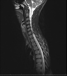
Figure 2: Day 1 Attempted IR Ablation with loss of MEPs to Left Lower Extremity

Figure 3: Pre-embolization fluoroscopy of Spinal Cord AVM
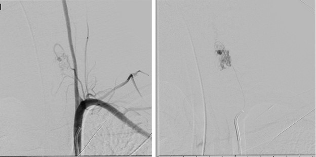
Figure 4: Day 2 IR lidocaine provocation challenge decreased amplitude and increased latency.

Figure 5: Post-embolization fluoroscopy of Spinal Cord AVM.
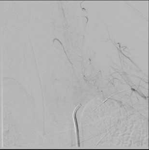
Table 1: Common Anesthetics and the effects on SSEPs and MEPs5. (↓ decrease, ↑ increase, ↔ minimal or no change)
| Anesthetic Agent | SSEP | MEP | ||
| Amplitude | Latency | Amplitude | Latency | |
| Propofol | ↓ | ↑ | ↓ | ↑ |
| Opioids | ↔ | ↔ | ↔ | ↔ |
| Etomidate | ↑ | ↑ | ↑ | ↔ |
| Ketamine | ↑ | ↑ | ↑ | ↔ |
| Benzodiazepines (low dose -premedication) | ↔ | ↔ | ↔ | ↔ |
| Barbiturates | ↓ | ↑ | ↓ | ↑ |
| Dexmedetomidine | ↓ | ↔ | ↓ | ↔ |
| Inhalation Anesthetics | ↓ | ↑ | ↓ | ↑ |
| N2O | ↓ | ↑ | ↓ | ↑ |